





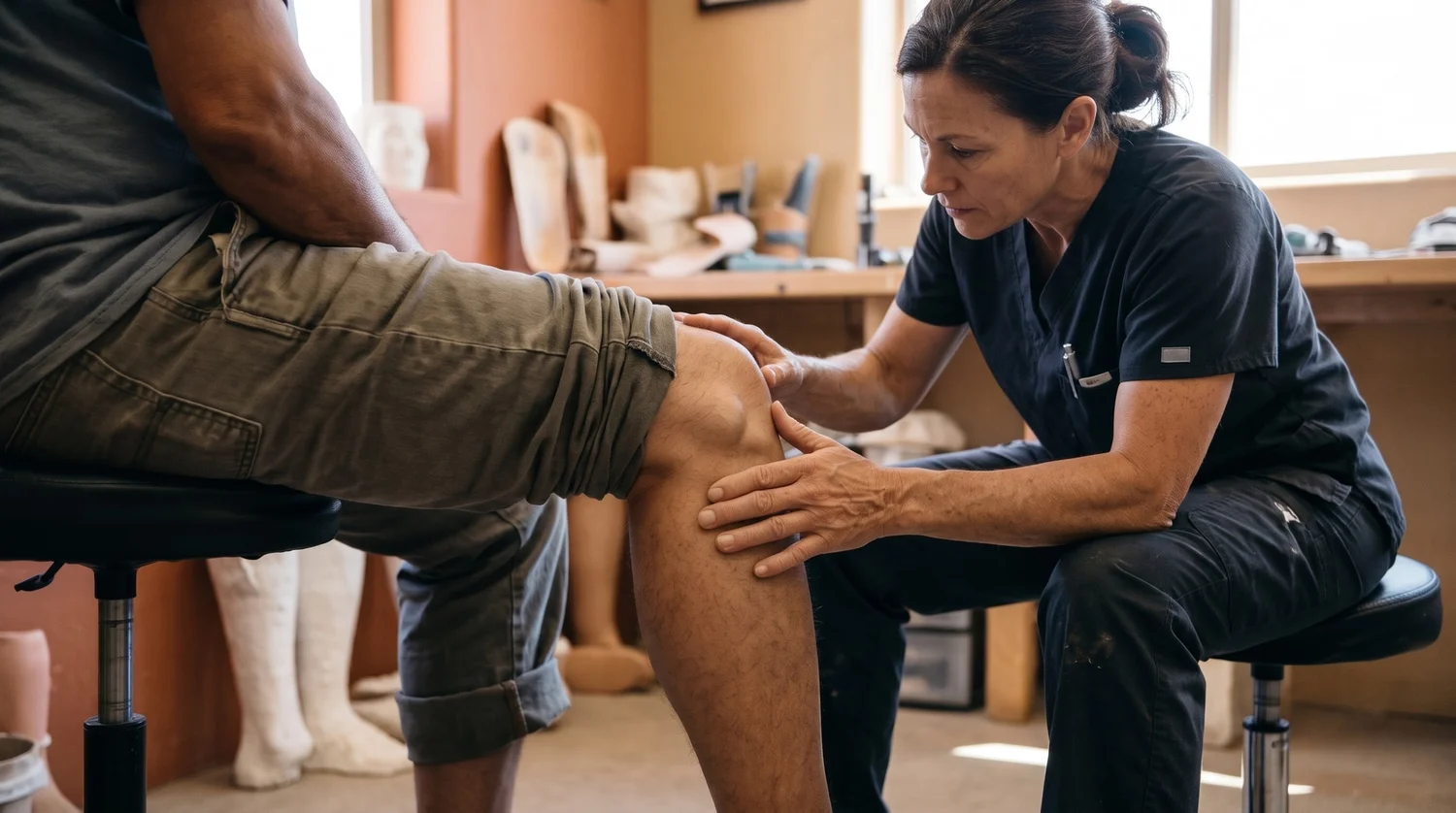
Most popliteal aneurysms cause no symptoms and are discovered incidentally during imaging for another condition. However, this bulge behind the knee can become dangerous if a clot blocks blood flow to the lower leg, potentially threatening the limb. PrimeCare provides prosthetic care and rehabilitation after limb loss, serving Las Cruces, Albuquerque, and Ruidoso.
What Is a Popliteal Aneurysm?
A popliteal aneurysm is a localized widening of the popliteal artery, the vessel that runs behind the knee and carries blood to the lower leg. Doctors generally define it as a diameter greater than about 1.5 cm, or 50% wider than the artery should be. Unlike an aortic aneurysm, which poses a risk of rupture, a popliteal aneurysm more often causes problems when clots form inside it, blocking the artery or breaking loose and traveling farther down the leg.
The bulge can be smooth and spread along the vessel (fusiform) or pouch out to one side (saccular). Because the popliteal artery lies close to the tibial nerve and popliteal vein, an enlarging aneurysm can press on these structures, causing leg pain or swelling.

How Serious Is a Popliteal Aneurysm?
A popliteal aneurysm is a limb-threatening condition even though it rarely bursts. The real risk comes from clot formation inside the bulge. When a clot blocks the artery or showers debris into the smaller vessels of the lower leg, blood flow can stop suddenly. Patients who develop acute thrombosis or embolism have a relatively poor prognosis, with roughly a 15% amputation rate due to occlusion of the runoff vessels.
How Common Is a Popliteal Aneurysm?
Popliteal aneurysms are uncommon overall but are among the most common peripheral artery aneurysms, affecting the arteries in the arms and legs:
- They make up about 85% of peripheral aneurysms, making them the most frequently found aneurysm outside the aorta.
- They are bilateral, present in both legs, in roughly half of cases, so an aneurysm in one knee prompts a check of the other.
- They are associated with abdominal aortic aneurysms in about 40% to 50% of patients, which is why doctors often screen the abdominal aorta as well.
- Male sex and advanced age are the primary non-modifiable risk factors, with the vast majority of cases occurring in men and diagnosis peaking in their 60s and 70s.
How Rare Is a Popliteal Vein Aneurysm?
While popliteal artery aneurysms account for the majority of peripheral aneurysms, a popliteal vein aneurysm is extremely rare, detected in 0.1% to 0.2% of routine vascular ultrasound examinations. Unlike arterial aneurysms, which threaten the limb through local thrombosis and ischemia, a popliteal vein aneurysm carries a different major risk: breaking off clots that travel to the lungs, causing a potentially fatal pulmonary embolism (PE). Because treatment differs significantly depending on which vessel is affected, targeted vascular ultrasound is needed to determine whether the artery or vein is involved.
What Are the Symptoms of a Popliteal Artery Aneurysm?
Many popliteal aneurysms cause no symptoms and are found by chance during an exam or imaging for another reason. When symptoms appear, they usually come from either compression of nearby structures or reduced blood flow: leg pain, numbness, calf swelling, a feeling of fullness behind the knee, or calf cramping when walking. A pulsating lump behind the knee is the most recognizable sign.

Is a Pulse Behind the Knee Normal or a Warning Sign?
A faint pulse behind the knee can be normal, since the popliteal artery runs close to the surface there. What raises concern is a pulse that feels enlarged, throbbing, or like a distinct lump you can feel with the knee slightly bent. Anyone who notices a prominent pulsating mass behind the knee should have it evaluated by a physician, ideally with an ultrasound.
Sudden severe leg pain, a cold or pale foot, numbness, or loss of the foot pulse can signal that the artery has clotted off. That is a medical emergency. Call 911 or go to the nearest emergency room right away. Prompt treatment is critical because restoring blood flow quickly can help prevent limb loss.
Popliteal Aneurysm Compared with a Baker's Cyst
A soft lump behind the knee is often mistaken for a Baker's cyst, a fluid-filled swelling that is far more common and not dangerous. A few differences help tell them apart:
- A Baker's cyst feels soft and does not pulse; a popliteal aneurysm often has a throbbing quality that matches the heartbeat.
- A cyst is filled with joint fluid, while an aneurysm carries flowing blood, so imaging that shows blood flow (a duplex ultrasound) distinguishes the two.
- A thrombosed aneurysm can lose its pulse and look like a firm cyst on a basic scan, which is why a vascular-focused ultrasound matters when there's any doubt.
Causes and Risk Factors
Most popliteal aneurysms develop from atherosclerosis, the same wall-weakening process behind many aortic aneurysms. Over time, the artery wall loses strength and begins to stretch under normal blood pressure. Less commonly, popliteal aneurysms are linked to inflammatory blood vessel diseases, connective tissue disorders such as Marfan or Ehlers-Danlos syndrome, or repeated mechanical stress around the knee. Atherosclerosis also drives peripheral artery disease, a related circulation problem that can put the lower limb at risk.
Who Is Most at Risk?
Certain factors show up repeatedly in people diagnosed with a popliteal aneurysm:
- Male sex, which accounts for the large majority of cases
- Age over 50 to 60
- A long history of smoking
- High blood pressure and high cholesterol
- A personal or family history of aneurysms or cardiovascular disease
How a Popliteal Aneurysm Is Diagnosed
Because a physical exam alone can miss an aneurysm — especially one filled with clot — imaging confirms the diagnosis. Duplex ultrasound is the usual first test: it measures the artery, shows blood flow, and can be repeated safely over time. When planning surgery, a CT angiogram or MR angiogram provides detailed images of the artery and the blood vessels farther down the leg. Since these aneurysms travel in company, a diagnosis often leads to a check of the other leg and the abdominal aorta.
Treatment Options for Popliteal Aneurysm
A vascular surgery team decides on the treatment, basing it on the aneurysm's size, whether it contains clot, and whether it's causing symptoms. Small, asymptomatic aneurysms may be monitored over time, while larger or symptomatic ones are typically repaired to reduce the risk of clotting and limb loss.
When Is Surgery Recommended?
General guidance from vascular surgery bodies points to repair in these situations:
- An asymptomatic aneurysm larger than about 2 cm, which is treated electively to avoid limb-threatening ischemia;
- Any aneurysm causing symptoms, regardless of size;
- An aneurysm containing significant clot or one that is compressing the nerve or vein behind the knee;
- Aneurysms smaller than 2 cm without symptoms, which are usually followed with regular ultrasound surveillance rather than surgery.
Open Surgical Repair or Endovascular Stenting
Two main approaches exist. Open surgery bypasses or excludes the aneurysm, frequently using the patient's own saphenous vein as the graft. The endovascular approach places a stent graft inside the artery through a small puncture. Both have a role, but the knee is a joint that bends thousands of times a day, and stent grafts placed across it face higher early re-intervention and clotting rates than open vein bypass. The right approach depends on the patient's anatomy and overall health, with a vascular surgeon helping determine the most appropriate treatment.
When a Popliteal Aneurysm Leads to Amputation
Most people with a popliteal aneurysm never lose a limb, especially when the problem is caught and treated early. Amputation becomes necessary in a minority of cases — typically after a sudden clot has cut off blood flow long enough that the tissue below can't recover, or when infection or gangrene has set in. In that setting, amputation can be a limb-preserving and often life-saving procedure that removes damaged tissue, controls pain and infection, and opens the door to prosthetic rehabilitation and a return to walking.
Below-Knee Compared with Above-Knee Amputation
The level of amputation depends on how much healthy circulation remains:
- A below-knee (transtibial) amputation is possible when blood flow through the thigh is intact. Keeping the natural knee joint is the single biggest factor in how quickly and easily someone adapts to a prosthetic leg.
- An above-knee (transfemoral) amputation is needed when clotting extends higher into the thigh vessels and the knee can't be preserved. A prosthetic knee then takes over that function.
Preserving the natural knee joint directly influences the biomechanics of walking and the prosthetic technology needed to restore your gait, as shown in the roadmap below:
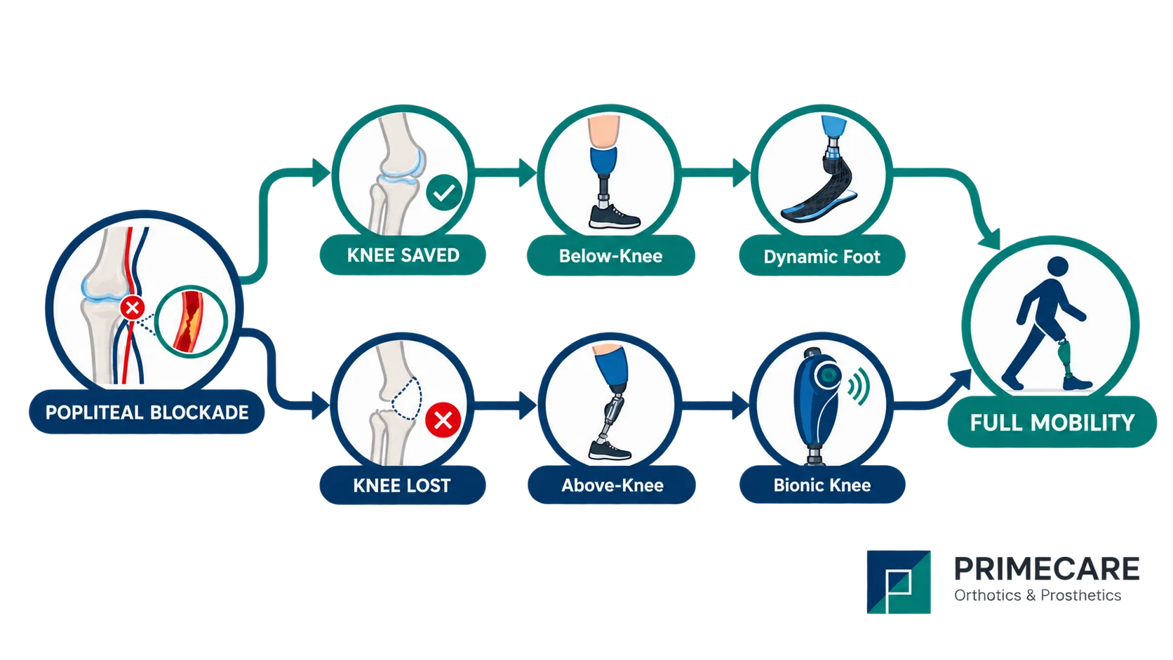
Figure 1: Prosthetic care pathways following popliteal occlusion. Preserving the knee joint allows for a transtibial prosthesis with a dynamic foot, while higher arterial blockages often utilize a transfemoral system with a microprocessor bionic knee.
Prosthetic Care After a Popliteal Aneurysm Amputation
This is where PrimeCare comes in. After the surgical team completes an amputation and the residual limb begins to heal, a prosthetist designs and fits the lower-limb prosthesis that gets you standing and walking again. The same prosthetic care applies whether limb loss comes from an aneurysm, traumatic amputation, or another cause, and PrimeCare works with people across New Mexico through each stage of it.
Below-Knee Prosthetics
For a transtibial amputation, a below-knee prosthesis pairs a custom socket with a pylon and a dynamic prosthetic ankle or foot system. Because the natural knee remains, many patients regain steady, confident walking.
Above-Knee Prosthetics
For a transfemoral amputation, an above-knee prosthesis incorporates an advanced prosthetic knee into the system, with component selection tailored to align with each person's mobility goals.
Rehabilitation After Limb Loss
Getting back on your feet involves several stages of prosthetic fitting, rather than just a single appointment. The process begins with residual limb shaping and compression, followed by casting or scanning for a custom socket. From there, it progresses to diagnostic fitting, alignment, and gait training. Follow-up visits then fine-tune the fit as the limb changes.